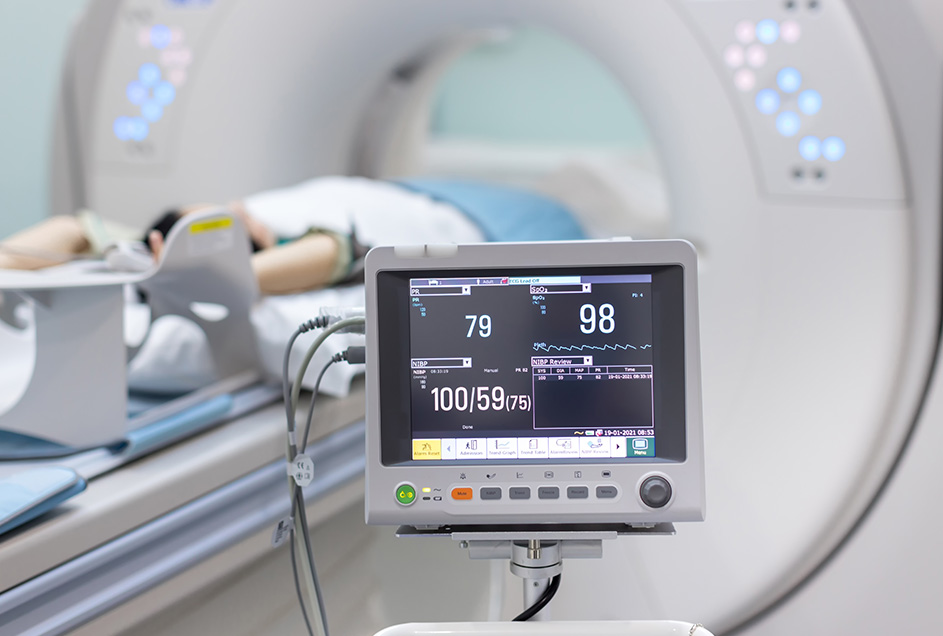
Advanced cardiac imaging describes techniques specifically designed to assess how well your heart is working. Combined with the results of other tests where necessary, this insight allows your cardiologist to assess your risk, confirm a diagnosis, and guide treatment recommendations.

CTCA is an imaging technique used to evaluate the condition of the arteries supplying your heart muscle, known as the coronary arteries. CTCA is sometimes referred to as a cardiac CT scan or coronary CT angiogram. Compared to a conventional or invasive angiogram, the CTCA is less invasive and faster while still carrying high diagnostic value when used for the right patients.
With the use of a contrast dye injected into a vein, a CT coronary angiography scan highlights the space through your coronary arteries and allows your cardiologist to see how well blood is flowing through these vessels. This can highlight any blockages, including soft and calcified plaque, revealing the location and extent of these obstructions. A cardiac CT angiogram can also show changes to the heart muscle, the valves, the membrane surrounding the heart (pericardium), and the chest.
CTCA is very useful in identifying individuals with coronary artery disease, and can also be used to check whether a coronary bypass graft is working as expected in select cases.
A CTCA is performed as a day procedure at a radiology practice and then interpreted and reported by our cardiac imaging subspecialist.
It may be recommended if you have:
Though the duration of the actual scan is only about 20 minutes, your appointment can take around 2-3 hours in total. The best results are obtained with a low heart rate at the time of scanning. You are advised to not consume caffeine within 24 hours of the scan, and may be advised to take pre-medication like beta blockers to slow the heart.
Also known as a calcium score test, a CAC scan is a non-invasive computed tomography (CT) scan performed without intravenous contrast, designed to evaluate the degree of calcified plaque in your arteries. This produces a coronary calcium score score, which indicates your risk of heart attack or cardiovascular disease over the next 10 years. A calcium score test has demonstrated value in predicting heart attack risk particularly in asymptomatic individuals at an intermediate risk of cardiovascular disease.
Calcified plaque in the coronary arteries is a direct marker of atherosclerosis and the hardening of these blood vessels. A CAC score can fall anywhere from zero to over 400. The higher your CAC score, the more extensive the amount of plaque and the higher your risk of coronary artery disease.
The calcium score test is a type of cardiac CT. Unlike a CT coronary angiography, a calcium score test is entirely non-invasive and does not require the use of contrast dye.
You may be recommended to undergo CAC scoring if you:
The results of your CAC scan give your cardiologist more guidance in tailoring a treatment plan to you. This may involve reassurance that your risk is low and preventative medicine is not required, or that intervention is needed promptly to prevent a heart attack.
A conventional, invasive coronary angiogram provides a visual of the blood flow through your heart’s blood vessels, and is used to identify areas of narrowing or blockage that may increase your risk of a cardiac event.
Where CTCA injects the contrast dye via an IV line, the invasive coronary angiogram requires a catheter to be inserted into your coronary artery. Access is generally gained via either the groin or the wrist, with local anaesthetic given. Although this type of coronary angiogram is more invasive and does come with greater risks compared to CTCA, it can provide a higher level of detail and may be more accurate in detecting problems in high-risk patients. Another significant advantage of a conventional coronary angiogram is that your cardiologist can treat the narrowing or obstruction of your arteries at the same time as detecting them, reducing your need for an additional procedure.
Invasive coronary angiography can be performed by one of our interventional cardiologists. You may be recommended an ICA if:
A cardiac MRI scan uses magnetic fields and radio waves to create images of your heart and associated blood vessels. It is a non-invasive imaging procedure, providing structural and functional details about your heart for the evaluation of many different cardiovascular conditions.
There is no ionising radiation used in MRI, but due to the high-powered magnets in the machine, you must remove all metal objects from your person, such as watches and jewellery. If you have a pacemaker or other implanted device, an MRI may not be appropriate for you.
Contrast dye is injected intravenously during your scan, and you will also have an ECG recorded simultaneously.
A cardiac MRI scan may be ordered and interpreted by our cardiac imaging subspecialist, and takes about 45 minutes to an hour to complete. A cardiac MRI scan may be recommended to investigate:
At Harbourside Cardiology, our team is dedicated to providing holistic, patient-centred care that aligns with the most recent clinical guidelines and research. We are with you every step of your health journey, providing continuity of care of all heart conditions from initial assessment through to treatment and recovery.